Eye Tests
Eye Tests
There are lots of different kinds of eye tests, and not everyone will need each kind. It will depend on your health and your risks for certain eye conditions.
Why have an eye test?
If you notice any symptoms with your eyes, or any changes to your vision, you should see an optometrist or doctor to organise an eye test. Regular eye tests can detect and diagnose eye problems in the early stages.
Common Eye Tests
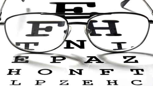
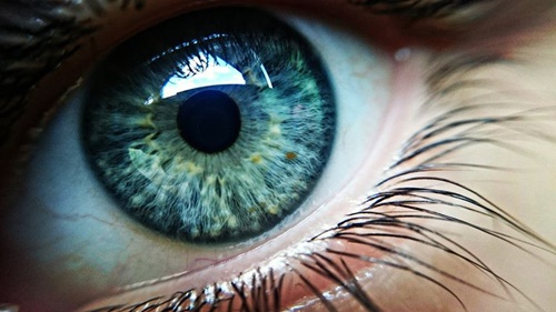
Visual Acuity Testing
A visual acuity test is an eye exam that checks how well you see the details of a letter or symbol from a specific distance. Visual acuity refers to your ability to discern the shapes and details of the things you see. Others include color vision, peripheral vision, and depth perception.
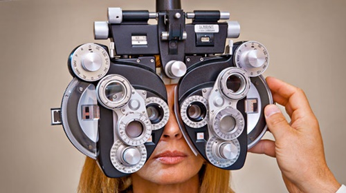
Refraction
A refraction test is what the doctor uses to get your eyeglasses prescription. You look at a chart, usually 20 feet away, or in a mirror that makes things look like they’re 20 feet away. You’ll look through a tool called a phoropter. It lets the doctor move lenses of different strengths in front of your eyes.
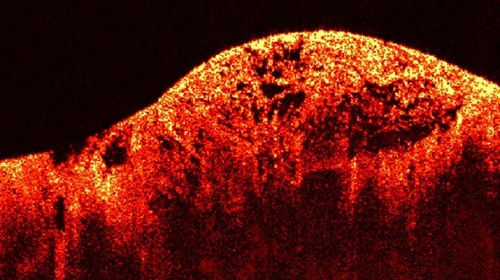
Optical Coherence Tomography
Optical Coherence Tomography gives an unprecedented view of the macula and optic nerve. The contour maps and “virtual” cross section of the tissues gives invaluable information of eye health. This includes early detection of eye conditions such as macula degeneration and glaucoma. Stunning detail of the retinal structures, and a big step up from retinal photography.
Corneal Pachymetry
Corneal ultrasound pachymetry allows easy measurement of corneal thickness. Using this technology we can make a more accurate assessment of glaucoma risk – thinner corneas are more of a concern. It gives a more accurate understanding of what the real eye pressures are and the need for treatment.
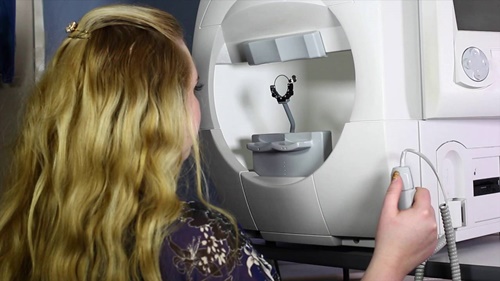
Computerised Visual Field
Peripheral vision testing is important in the screening for optic nerve abnormalities such as glaucoma. Subtle defects in side vision occur as the optic nerve fibres deteriorate. Most people are not consciously aware of these changes until the disease is well advanced. Visual field assessment is another part of the puzzle for glaucoma diagnosis.
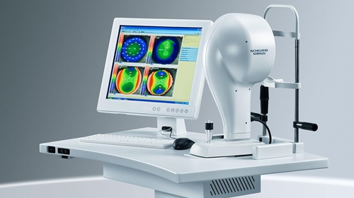
Corneal Topography
Mapping the shape and curvature of the cornea is useful in: measuring the amount of regular and irregular corneal astigmatism, advanced contact lens fitting, assessing suitability for laser surgery and diagnosing eye disease such as Keratoconus.
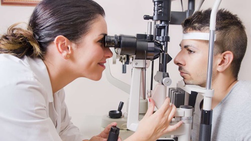
Applanation Tonometry
This test measures fluid pressure in your eye. The test involves using a slit lamp equipped with forehead and chin supports and a tiny, flat-tipped cone that gently comes into contact with your cornea. The test measures the amount of force needed to temporarily flatten a part of your cornea.
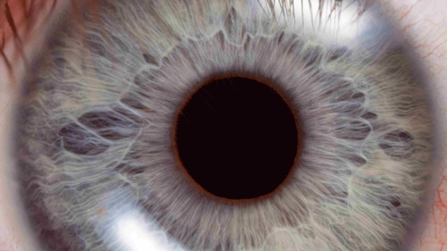
Dilated Pupillary Exam
Pupil dilation is performed to purposefully increase the size of the pupils during an eye exam so that the eye doctor can fully examine the health of the optic nerve and retina. The exam is critical to preventing and treating eye conditions that could potentially lead to vision loss.
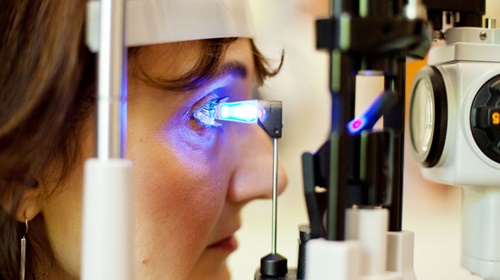
Slit Lamp Exam
Your eye doctor may use a microscope called a slit lamp to examine the front of your eye. The microscope focuses an intense narrow line of light on your eye. The slit lamp provides a magnified, 3D view of the eye and allows your doctor to detect any small abnormalities. Used with special lenses held close to the eye, the slit lamp also provides detailed views of the back of the eye.